Define glycolysis and describe its process
Glycolysis is the first stage of cellular glucose catabolism. Skeletal muscle is the largest tissue containing enzymes for glycolysis. Basically, this is the breakdown of a 6-carbon glucose molecule into 2x 3-carbon carboxylic acid pyruvate molecules, finally into lactate via the enzyme lactate dehydrogenase. Both the pyruvate and lactate anions have a carboxyl group. Moving from glucose to pyruvate creates a proton. The process of moving from pyruvate to a lactate anion consumes a proton, making this a buffering event, helping to control for pH changes. Lactate dehydrogenase catalyzes the conversion of pyruvate anion to lactate anion. This reaction has a high standard free energy change and equilibrium constant.
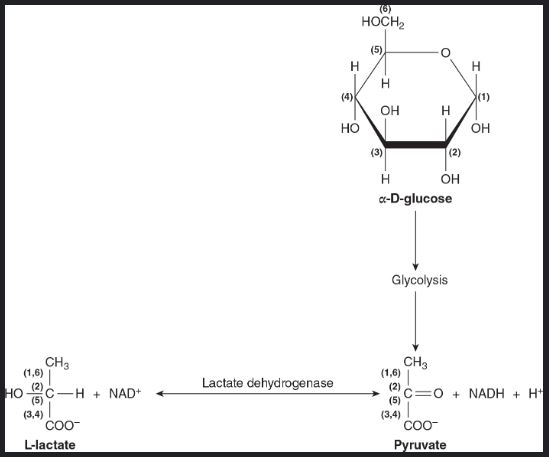
Glycolysis occurs in the cytosol of cells where the glycolytic enzymes are found. Glycolysis operates in both slow and rapid pathways depending on the flux of metabolism. The figure below illustrates (A) slow glycolysis and (B) fast glycolysis.
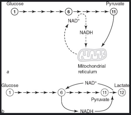
While glycolysis occurs often in 12 steps, it is helpful to break it down into two big six-part steps. The first step ends with the aldolase and isomerase steps. The regulatory enzyme glyceraldehyde 3-phosphate dehydrogenase kicks off the second of the six steps leading to the formation of lactate anion. The first step is regulated by the two enzymes phosphofructokinase and hexokinase. The second step is catalyzed by 3-phosphoglycerate kinase and pyruvate kinase to phosphorylate ADP to ATP. The first part consumes 2 ATP and the second part produces 4 ATP, meaning glycolysis has a net yield of 2 ATP/glucose. The abundance of glycolytic enzymes in muscle tissue leads to many of these reactions happening readily for short bursts of power.
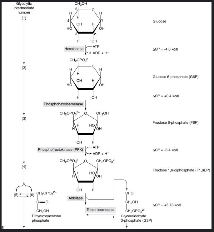
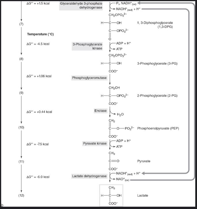
For emphasis: Glycolysis does not make lactic acid. It makes lactate anion from pyruvic acid (pyruvate and a proton). Lactate formation is a acid-buffering step.

What is the role of the liver in the supply of glucose to an exercising person?
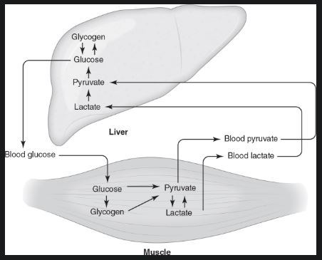
The liver maintains blood glucose by breaking down glycogen and performing gluconeogenesis, supplying glucose to active muscles. Gluconeogenesis is the creation of new glucose from precursor molecules, mainly lactate.
Glucose from the duodenum moves to the liver through the hepatic portal vein. The textbook gives the example that after a 75 gram glucose tolerance test, 80% is held by the liver as glycogen; after 30 minutes the liver will release this in the form of glucose over several hours. The liver functions to maintain euglycemia; meaning, it regulates blood glucose homeostasis. One of the most carefully controlled physiologic variables is the concentration of glucose in blood plasma (have you thanked your liver recently?). Much of this liver released glucose heads for skeletal muscle where it is stored as glycogen to be converted to lactate via cellular metabolism. This lactate then enters the blood stream back to the liver for the process to begin again.
From the energetic demand of exercise, the supply of blood glucose is challenged. The liver then needs to churn out more glucose to help maintain euglycemia. Exercise at 50% of VO2 max liver glucose production (called hepatic glucose output) will approximately rise to 3.5 mg/kg x min, from 1.8 mg/kg x min at rest.
What discovery about mitochondria led to the intracellular lactate shuttle concept?
The discovery that liver, heart, and skeletal muscle mitochondria oxidizes lactate at higher rates than pyruvate sparked the discovery of the intracellular lactate shuttle concept. When cytosolic lactate concentrations rise, lactate shuttled to nearby mitochondria is oxidized. The production of lactate through glycolysis was thought to be a dead end metabolite. Now it is understood that lactate is the link between glycolytic and oxidative metabolisms. This shuttle concept helps us understand oxidative glycolysis, when fully oxygenated cells consume glucose and produce lactate.
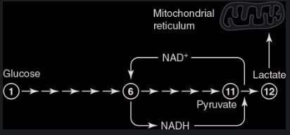
The intracellular lactate shuttle supports aerobic glycolysis. Rapid glycolysis can occur resulting in the accumulation of lactate because of the simultaneous high rate of oxygen consumption in mitochondria.
Describe the enteric and systemic phases of the postprandial lactate shuttle
The postprandial lactate shuttle (PLS) was discovered in 2024. Robert Leija et al. from the Brooks lab at the University of California, Berkeley demonstrated that the first metabolite to appear in arterial blood after eating is lactate, not glucose. They established that the liver prefers to make glycogen from lactate, not glucose. This shuttle functions to distribute dietary carbohydrate carbons. From the textbook: “Glycolysis leading to lactate production is not a “muscle thing, it’s an “everything”. “
There are two phases of the postprandial lactate shuttle. The enteric phase is described as when the duodenal enteric cells convert glucose to lactate, this lactate then travels via the hepatic portal vein, hepatic veins, inferior vena cava, then into systemic circulation, marking the second phase of the PLS, where lactate is distributed via hepatic glucose release. This research helped to establish that lactate production is not the result of skeletal muscle hypoxia, but the end product of glycolysis in fully aerobic tissue. The PLS is a vehicle of delivering lactate throughout the body.
Red skeletal muscle and the Cori cycle
Red skeletal muscle is red because of its myoglobin density, (i.e. high capacity for lactate oxidation). This muscle tissue is often referred to as slow-twitch skeletal muscle, but is better defined as slow-oxidative (SO Type I). White muscle fibers are fast glycolytic (FG Type IIb). FG Type IIb cells can shuttle lactate produced to the SO type I fibers for oxidation and energy production. This is known as the cell-cell lactate shuttle.
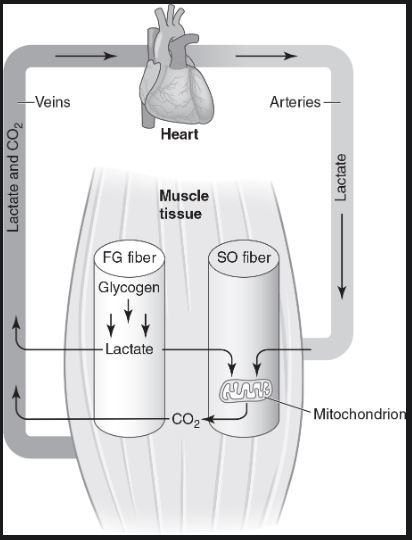
When lactate accumulation exceeds the cells ability to oxidize it, the lactate is then brought to the liver to be converted into glucose in a process called the Cori cycle. The body is able to create glucose from the byproducts of glycolysis termed gluconeogenesis.
Brooks, G. A., Fahey, T. D., & Baldwin, K. M. (2026). Exercise Physiology: Human Bioenergetics and Its Applications (6th ed.). Human Kinetics. [1, 2]